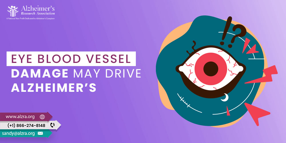
A new study revealed that abnormalities in the eye blood vessels could aid in the early detection of Alzheimer’s. The team identified an aberration in the blood-retinal barrier, which stops hazardous compounds from entering the retinal tissue, by comparing the retinal blood vessels of Alzheimer’s patients, people with mild cognitive impairment, and healthy volunteers.
According to a study by Cedars-Sinai researchers published in the journal Alzheimer’s & Dementia, anomalies in the blood vessels in the eye play a significant role in the development of Alzheimer’s. These modifications correspond to brain changes, opening a new avenue for early disease detection.
Maya Koronyo-Hamaoui, the senior author, stated that this research offers fresh insight into the vascular abnormalities linked to Alzheimer’s disease, particularly in the retina, the layer of nerve tissue in the back of the eye. Additionally, it highlights the damage that Alzheimer’s does to the blood vessels in the retina, opening up a novel, noninvasive route to early detection and disease progression monitoring.
The Research Findings
Researchers evaluated the blood vessels in the retinas of 27 donors with normal cognition, 10 with MCI (mild cognitive impairment), and 24 with Alzheimer’s.
They discovered one of the earliest symptoms of Alzheimer’s disease in participants with Alzheimer’s and MCI: the breakdown of the blood-retinal barrier, composed of closely bound cells that keep hazardous substances from penetrating the retinal tissue.
The researchers observed a loss of up to 70% in that barrier in Alzheimer’s patients, implying that toxic substances can flow through and penetrate the retinal tissue.
Damage to the blood-retinal barrier has been linked to different types of vascular illness in the brain as well as the condition known as cerebral amyloid angiopathy (CAA), which involves the amyloid proteins buildup in small blood vessels.
According to Koronyo-Hamaoui, post-mortem brain tissue samples are the only current means to identify CAA in patients. With further research and the development of enhanced retinal imaging technology, vascular and blood-retinal barrier impairment may provide the first opportunity to diagnose CAA in living individuals.
The study also discovered that amyloid beta 40 protein deposits built up in the retinal arteries of people with Alzheimer’s, stiffening the arteries, interfering with blood flow, and preventing the vessels from removing dangerous compounds from the retina. Further research is required to identify whether the deposits only accumulate as a result of blood vessel injury or actually contribute to the damages.
Rich in blood vessels, retinal and brain tissues require enough blood flow for optimal function. The restriction of blood supply, which may occur due to the damage, means these cells do not receive the oxygen and nutrients they require. Advanced retinal imaging is under development, but the Food and Drug Administration has not yet given it the go-ahead. It would allow noninvasive examination of blood vessels and protein buildup in living patients.
What did the researchers conclude?
Keith L. Black, MD, chair of the Department of Neurosurgery and the Ruth and Lawrence Harvey Chair in Neuroscience at Cedars-Sinai, stated that the retina had received extensive attention since it is a central nervous system window and an anatomical extension of the brain. This research contributes to current advances in sophisticated retinal imaging and the development of other retinal biomarkers to enhance the science of Alzheimer’s early detectionprogression of Alzheimer’s. Therefore, Koronyo-Hamaoui urges people to do everything they can to maintain a healthy circulatory system, particularly blood vessels in the retina and brain, to help stave off dementia and CAA.
Controlling hypertension, eating a low-sugar diet, limiting alcohol use, and quitting smoking all assist in preventing chronic inflammation and blood vessel damage.
References
- Shi, H., Koronyo, Y., Fuchs, D.T., Sheyn, J., Jallow, O., Mandalia, K., Graham, S.L., Gupta, V.K., Mirzaei, M., Kramerov, A.A. and Ljubimov, A.V., 2023. Retinal arterial Aβ40 deposition is linked with tight junction loss and cerebral amyloid angiopathy in MCI and AD patients. Alzheimer’s & Dementia.
- Study: Blood Vessel Damage could be an Alzheimer’s Driver. Cedars Sinai. https://www.cedars-sinai.org/newsroom/study-blood-vessel-damage-could-be-an-alzheimers-driver. Published Online: 19th May, 2023. Accessed: 2nd June, 2023.
- Blood Vessel in the Eye May Drive Alzheimer’s Disease. Technology Networks. https://www.technologynetworks.com/neuroscience/news/blood-vessel-damage-in-the-eye-may-drive-alzheimers-disease-373622. Published Online: 22nd May, 2023. Accessed: 2nd June, 2023.
- Eye Blood Vessel Changes: A New Window into Alzheimer’s Diagnosis. Neuroscience. https://neurosciencenews.com/alzheimers-eye-blood-vessels-23289/. Published Online: 19th May, 2023. Accessed: 2nd June, 2023.